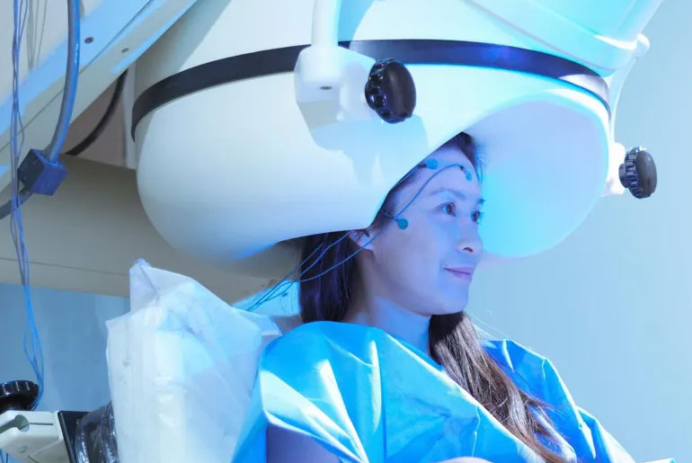
Researchers at Simon Fraser University are employing a novel kind of brain imaging that may enhance the way medications are given to treat Parkinson’s disease.
The new study examines why levodopa, the primary medication used in dopamine replacement treatment, can occasionally be less successful in individuals. It was published in the journal Movement Disorders.
Usually, the medication is recommended to assist lessen the neurodegenerative disease’s movement symptoms.
Although the great majority of people find that it helps them with their problems, not everyone benefits equally.
To understand why, an SFU partnership with Swedish researchers has using magnetoencephalography (MEG) equipment to assess the drug’s impact on brain impulses.
“Parkinson’s is the second most prevalent neurodegenerative disease worldwide and it is the most rapidly increasing, in terms of incidence,” states Alex Wiesman, assistant professor in biomedical physiology and kinesiology at SFU.
“Treating this disease, both in terms of helping people with their symptoms, but also trying to find ways to reverse the effects, is becoming more and more important.”
“If clinicians can see how levodopa activates certain parts of the brain in a patient, it can help to inform a more personalised approach to treatment.”
Researchers from Sweden’s Karolinska Institute collaborated on the study, gathering data from a very small sample of 17 Parkinson’s disease patients using MEG.
To determine how and where the treatment affected brain activity, researchers mapped the subjects’ brain signals before and after taking the medication.
MEG is a cutting-edge non-invasive technique that detects the magnetic fields generated by electrical signals in the brain.
It can support the study of brain illnesses and disorders, including as brain injuries, tumors, epilepsy, autism, mental illness, and more, by researchers and physicians.
Wiesman and colleagues created a novel methodology that enables them to “search” the brain for off-target medication effects using this uncommon brain imaging technology.
“With this new way of analyzing brain imaging data, we can track in real time whether or not the drug is affecting the right brain regions and helping patients to manage their symptoms,” states Wiesman.
“What we found was that there’s sometimes ‘off target’ effects of the drug. In other words, we could see the drug activating brain regions we don’t want to be activating and that’s getting in the way of the helpful effects.”
“We found that those people who showed ‘off target’ effects are still being helped by the drug, but not to the same extent as others.”
As a neurodegenerative condition, Parkinson’s disease causes gradual brain damage over time. It primarily impacts the dopamine-producing neurons in the substantia nigra, a particular region of the brain.
A variety of movement-related symptoms, including tremors, sluggish movement, stiffness, and balance issues, can be experienced by people with Parkinson’s disease.
Wiesman believes that a greater knowledge of how levodopa influences a person’s brain signals may lead to better prescription practices for Parkinson’s disease medications.
“This might be really helpful for tracking individualized responses to these types of drugs and helping with prescribing and therapeutics,” he states.
“So maybe we try different medications, maybe we adjust dosages differently. And this helps clinicians get at that question of how we prescribe personalized medicine in a way that really helps the patient.”
“The more we can personalize that approach, make it more expedient, make it a bit more specific to that person, the better.”
This new type of brain imaging analysis is not only for studying Parkinson’s disease; any medications that affect brain signaling can be studied using the method developed by Wiesman and colleagues.
SFU’s ImageTech Lab, at the Surrey Memorial Hospital, is home to the only MEG in western Canada.
“We have this really fantastic technology right here at SFU, and combined with the new analysis approaches that we’re developing, it gives us a really unprecedented look into what’s happening in the brain,” states Wiesman.
“We can use this technology moving forward to study Parkinson’s disease in ways that no one has ever done before worldwide.”
“Our next step is to take our new approach and apply it to a larger patient group. We also need to translate this research to more accessible brain imaging methods, like electroencephalogram (EEG).”
“Ultimately, we want to make sure this technology is useful for a diverse population and more widely accessible to patients with Parkinson’s disease.”
Disclaimer:
The information contained in this article is for educational and informational purposes only and is not intended as a health advice. We would ask you to consult a qualified professional or medical expert to gain additional knowledge before you choose to consume any product or perform any exercise.


